
Abdominal Anatomy Images: A Deep Dive Into The Core Of Human Anatomy
Have you ever wondered what's really going on beneath your skin in the abdominal region? If you're here, chances are you're looking to uncover the mysteries of abdominal anatomy images. Whether you're a student, a healthcare professional, or just someone curious about the human body, this article is your ultimate guide. Let's dive right into it, shall we?
Let’s be honest, understanding the abdominal anatomy can feel like trying to decode a complex map. But don’t worry—we’ve got your back (and your core)! Abdominal anatomy images are more than just pictures; they’re like roadmaps that help us understand how everything fits together in the abdominal cavity. Whether you're studying for an exam, preparing for surgery, or simply satisfying your curiosity, these visuals play a crucial role.
Before we get into the nitty-gritty, let’s set the stage. The abdomen is like the body’s control center, housing vital organs that keep us ticking. From the stomach to the intestines, every part plays a role in keeping our systems running smoothly. So, buckle up because we’re about to explore the fascinating world of abdominal anatomy images, and trust me, it’s going to be an eye-opening journey.
- Kathleen Hilfiger The Untold Story Of A Fashion Empires Heart And Soul
- Girls Showering A Comprehensive Guide To Understanding The Routine Benefits And Privacy
Why Abdominal Anatomy Images Matter
Abdominal anatomy images aren’t just for show—they’re tools that can make or break your understanding of the human body. Imagine being a doctor trying to diagnose a patient without a clear picture of what’s happening inside. Sounds tough, right? That’s where these images come in. They provide a visual representation of the complex structures within the abdomen, making it easier to identify issues and plan treatments.
For students, these images are like cheat sheets that help them ace their exams. For surgeons, they’re like blueprints that guide them through intricate procedures. And for the average person, they’re a way to understand their own bodies better. In short, abdominal anatomy images are the unsung heroes of healthcare.
How These Images Are Created
Ever wondered how those detailed abdominal anatomy images come to life? It’s not magic—it’s science! Most of these images are created using advanced imaging techniques like CT scans, MRIs, and ultrasounds. These technologies allow us to see inside the body without having to cut it open, which is a pretty big deal if you ask me.
- Kelly Albanese The Rising Star Of Modern Entertainment
- Anna Hall Hot The Rising Star You Need To Know About
Here’s a quick rundown of how it works:
- CT Scans: These use X-rays to create detailed cross-sectional images of the abdomen.
- MRIs: These use powerful magnets and radio waves to produce detailed images of soft tissues.
- Ultrasounds: These use sound waves to create images, often used for examining organs like the liver and kidneys.
Each method has its own strengths and is chosen based on what needs to be examined. Pretty cool, huh?
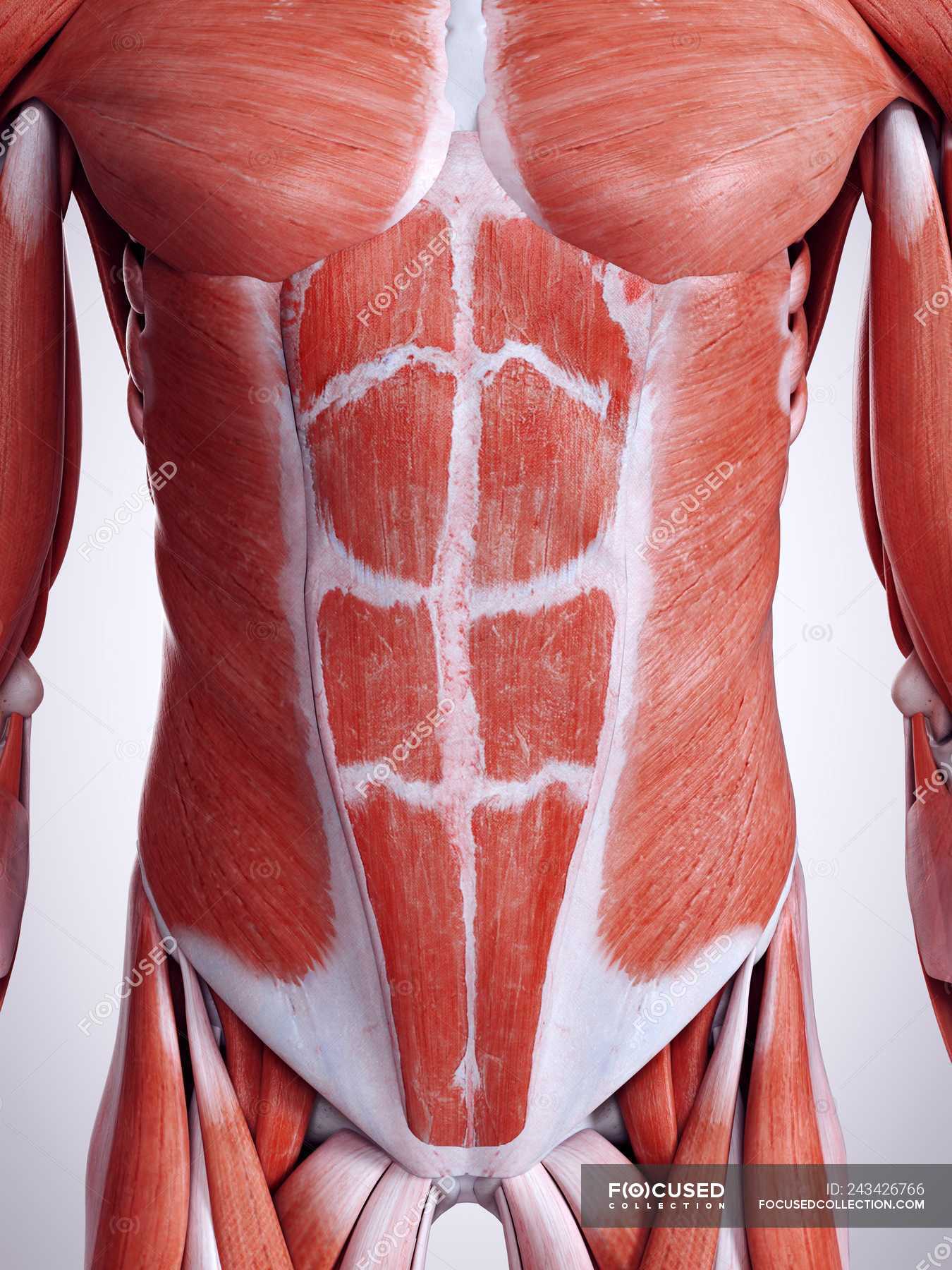
Key Organs in Abdominal Anatomy
Now that we know why abdominal anatomy images are important, let’s talk about the stars of the show—the organs. The abdomen is home to some of the most vital organs in the body, each with its own unique function. Here’s a quick look at some of the key players:
The Stomach
The stomach is like the body’s personal chef. It’s where food is broken down and prepared for digestion. Abdominal anatomy images often highlight the stomach’s location and structure, showing how it connects to the esophagus and small intestine. Without the stomach, digestion would be a mess!
The Liver
Think of the liver as the body’s chemical factory. It produces bile, processes nutrients, and detoxifies harmful substances. Abdominal anatomy images show the liver’s impressive size and position, often highlighting its lobes and blood vessels. It’s a powerhouse organ that deserves all the credit it gets.
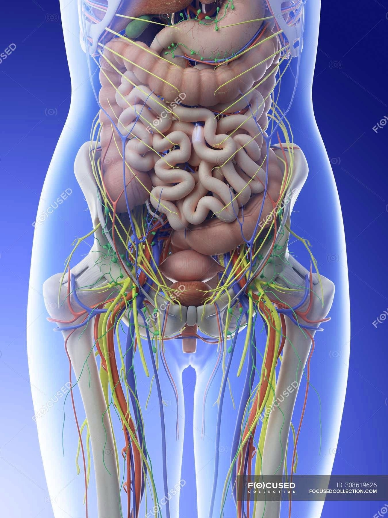
The Intestines
The intestines are like the body’s assembly line. They’re responsible for absorbing nutrients and moving waste out of the body. Abdominal anatomy images often depict the small and large intestines in great detail, showing how they twist and turn to fit inside the abdomen. It’s a fascinating process that keeps us fueled and functioning.
Common Abdominal Conditions
Unfortunately, the abdomen isn’t immune to problems. There are several common conditions that can affect the organs within. Let’s take a look at a few:
Gastroesophageal Reflux Disease (GERD)
GERD is a condition where stomach acid flows back into the esophagus, causing heartburn and other unpleasant symptoms. Abdominal anatomy images can help doctors identify issues with the lower esophageal sphincter, which is often the culprit in GERD cases.
Liver Disease
Liver disease can range from mild to severe, affecting the liver’s ability to function properly. Abdominal anatomy images are crucial in diagnosing conditions like cirrhosis and fatty liver disease, allowing doctors to plan effective treatments.
Intestinal Obstruction
An intestinal obstruction occurs when something blocks the intestines, preventing food and fluids from passing through. Abdominal anatomy images can help identify the cause and location of the obstruction, guiding doctors in their treatment decisions.
The Role of Abdominal Anatomy Images in Diagnosis
When it comes to diagnosing abdominal conditions, images are invaluable. They provide a clear picture of what’s happening inside the body, helping doctors make informed decisions. Whether it’s identifying a tumor, locating a blockage, or assessing organ damage, abdominal anatomy images are often the key to successful diagnosis.
Here’s how they’re used:
- Identifying Abnormalities: Images can highlight unusual growths or structures within the abdomen.
- Guiding Procedures: Surgeons rely on images to navigate during operations, ensuring precision and safety.
- Monitoring Progress: Images can track the progression of a condition over time, helping doctors adjust treatments as needed.
Without these images, diagnosing and treating abdominal conditions would be a lot more challenging. They’re truly the backbone of modern medicine.
Abdominal Anatomy Images in Education
For students and educators, abdominal anatomy images are more than just tools—they’re teaching aids. They help bring the complexities of the human body to life, making it easier for students to grasp difficult concepts. Whether it’s through textbooks, online resources, or interactive software, these images play a crucial role in education.
Here are a few ways they’re used in the classroom:
- Visual Learning: Images provide a visual representation of abstract concepts, aiding in comprehension.
- Interactive Tools: Many educational platforms offer interactive images that allow students to explore the abdomen in 3D.
- Real-World Applications: Students can see how these images are used in real-world medical scenarios, bridging the gap between theory and practice.
For anyone studying anatomy, these images are indispensable. They turn the abstract into the concrete, making learning more engaging and effective.
Trends in Abdominal Imaging Technology
Technology is constantly evolving, and the field of abdominal imaging is no exception. New advancements are being made all the time, offering even more detailed and accurate images of the abdomen. Let’s take a look at some of the latest trends:
3D Imaging
3D imaging is revolutionizing the way we view the abdomen. Instead of flat, two-dimensional images, we now have the ability to see organs in three dimensions. This provides a more comprehensive view, helping doctors make more accurate diagnoses.
AI in Imaging
Artificial intelligence is also making waves in the imaging world. AI algorithms can analyze images faster and more accurately than humans, identifying issues that might otherwise be missed. It’s like having a super-smart assistant in the room!
Portable Imaging Devices
Gone are the days of bulky imaging machines. Portable devices are becoming more common, allowing doctors to perform scans in the field or in remote locations. This is a game-changer for areas with limited access to healthcare.
Challenges in Using Abdominal Anatomy Images
While abdominal anatomy images are incredibly useful, they’re not without their challenges. Here are a few things to keep in mind:
Interpreting Images
Not everyone is trained to interpret medical images. Even experienced doctors can sometimes miss subtle details, which is why second opinions are often recommended. It’s important to have a trained professional analyze these images to ensure accuracy.
Cost and Accessibility
Advanced imaging technologies can be expensive, making them inaccessible to some patients. This is a challenge that healthcare systems around the world are working to address, but it remains a significant issue in many areas.
Patient Safety
Some imaging techniques, like CT scans, involve radiation exposure, which can pose risks to patients. It’s important to weigh the benefits against the risks when deciding whether to use these methods.
Conclusion
In conclusion, abdominal anatomy images are an invaluable resource in the world of healthcare. They help us understand the complexities of the human body, diagnose conditions, and educate the next generation of medical professionals. Whether you’re a student, a doctor, or just someone curious about the body, these images offer a fascinating glimpse into the inner workings of the abdomen.
So, the next time you come across an abdominal anatomy image, take a moment to appreciate the science and technology behind it. And if you’ve found this article helpful, don’t forget to share it with others who might benefit from the knowledge. After all, the more we know about our bodies, the better equipped we are to take care of them.
Table of Contents
Article Recommendations
- Kate Danson The Rising Star Taking The World By Storm
- Bending Over The Ultimate Guide To Mastering Flexibility And Strength

Detail Author:
- Name : Mr. Presley McClure
- Username : jgislason
- Email : joanie81@cummerata.net
- Birthdate : 2001-09-18
- Address : 3323 Emilie Locks Wilhelminefurt, CA 23592-3512
- Phone : (828) 949-4369
- Company : Krajcik and Sons
- Job : Offset Lithographic Press Operator
- Bio : Illum voluptatum et rerum aut itaque voluptatem dolores sunt. Modi itaque iste repellat rerum totam quis necessitatibus alias. Reiciendis in dolores reprehenderit quis.
Socials
linkedin:
- url : https://linkedin.com/in/ceasar.carroll
- username : ceasar.carroll
- bio : Expedita id earum ullam aliquid minima.
- followers : 3506
- following : 2714
instagram:
- url : https://instagram.com/ceasarcarroll
- username : ceasarcarroll
- bio : Dolor voluptatem impedit sit optio quam. Vitae quasi quia vero quos quidem.
- followers : 981
- following : 1456
